When monitoring pregnancy, “screening” is the most frequently mentioned topic, both by doctors and patients.
In order to clarify things, I would like to briefly explain the term “screening”. This term is used in many areas of daily living and means a fast and easily accessible method, by which from a general, unselected set of objects, people, documents, patients, etc. those that are special or different will be selected. In medicine, i.e. in obstetrics, this means that pregnant women who are different i.e. those with a condition that needs to be further examined are selected from general population.
There are several types of screening during pregnancy:
- ultrasound screening (the screening for Down syndrome is part of the ultrasound screening);
- hidden sugar screening;
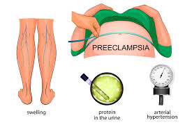
- high pressure screening and
- screening for premature delivery.
Of all the above, the term ultrasound screening is, of course, the most familiar to patients.
As someone who has been doing ultrasound in pregnant women for longer than 20 years, I want to clarify some terminological dilemmas that patients and even doctors often have.
Other terms are often used for ultrasound screening – 4D-screening, screening for anomalies, morphological ultrasound, Doppler-ultrasound, etc. I can say that all names are used for the same thing or term.
Ultrasound examination in certain specific periods of pregnancy. The ultrasound examination itself, i.e. the appropriate ultrasound machine, as a technical possibility has a 4D technique, Doppler or color Doppler. These additional features of ultrasound machines are not always necessary.
The use of these techniques depends on the doctor performing the examination. During the examination, the doctor himself decides whether he needs to include the appropriate technique.
There are also many misconceptions concerning the term or timing of the examination itself. In everyday practice, patients and doctors often insist that the examination must be performed within a period of one week and that it would not be feasible afterwards.
Another misconception!
It is true that there are certain specific periods (I will explain later why) when the examinations should be carried out, but the misconception is that it has to be in the 21st week (for the second screening) or the 12th week (for the first screening). The time frame ranges from 3 to 4 weeks, and even if it is not within the time frame, the examination can certainly be performed.
After all, for experienced doctors, every ultrasound examination of the fetus, in any part of the pregnancy, is in some way a screening.
It is generally accepted that there are three screenings during pregnancy.
I will now explain what is specific to each of them and why they are positioned in the appropriate periods of pregnancy.
Screening in the first trimester
With the completion of organogenesis, around the 12th week of gestation, as the name suggests, the formation of fetal organs is completed.
With modern high-resolution ultrasound machines, an initial morphological (anatomical) assessment of the fetus can already be made in this period. In other words, it means that all the organs of the fetus can be seen, as well as the behavior, i.e. fetal mobility. In this period, a number of possible disorders (defects) in the embryo can already be detected.
The time frame ranges from the 11th to the 14th week of gestation, although, in my experience, the period of full 12th or 13th week is ideal. During this time, the development of the embryo changes very quickly, so somewhere around the 13th week there is more information than in the 11th week.
As an integral part of the ultrasound examination in this period is the screening for Down syndrome.
Fetuses with Down syndrome, in most cases (85%), have certain characteristics in the first trimester. It must be noted that these are not morphological defects, but specific signs (markers), which these fetuses most often have (swelling of the neck, small nose, specific circulation).
It must be emphasized that even if the embryo has these characteristics, it does not necessarily mean that it has Down syndrome. It is just a signal that the embryo should continue to be monitored by other methods. As I explained above, the essence of screening is to single out those who require further testing.
To summarize once again.
- When the embryo has specific features of Down syndrome, additional tests must be
- performed to confirm the suspicion. Although rare, a normal embryo may have these
- characteristics, which are later lost.
- These specific manifestations (not defects) are lost later in pregnancy even in fetuses with
- Down syndrome. Therefore, this period of pregnancy is very important.
- Although very rare, there are embryos with Down syndrome that look completely normal,
- i.e. do not have the aforementioned features.
- For these reasons it must be reiterated that screening can detect a high percentage of
- fetuses with Down syndrome, but never 100%.
When screening for Down syndrome is discussed, the inevitable question that arises is the one about the biochemical screening, known as PRISCA.
Although the topic is ultrasound screening, I have to clarify certain misconceptions about this.
In many countries around the world, screening for Down syndrome consists of two methods – ultrasound and biochemical markers.
Biochemical markers are, in fact, hormones that are present in every pregnant patient. In patients carrying children with Down syndrome, the level of these hormones may be disturbed, i.e. become higher or lower. It should be noted that this does not occur in every patient who carries a fetus with Down syndrome. The most comprehensive world studies report that up to 60% of Down syndrome cases can be detected this way.
Today, a combination of ultrasound and biochemical markers is made, as the gold standard in the world, to reach the figure of 90% of prenatally detected embryos with Down syndrome.
How does it function in North Macedonia? What is PRISCA?
I would like to emphasize that the name PRISCA is not a name for biochemical screening. It is the name of the software (20 years old) that calculates the risk for Down syndrome. The software includes parameters such as the patient’s age, ultrasound parameters and hormone levels, i.e. biochemical parameters.
Here I must emphasize that, after more than 10 years of experience with this software, no satisfactory results are obtained, especially since we, doctors who monitor pregnancy, often have undefined results, confusing for both patients and us. Very frequently there are so-called false positive results, i.e. a result that shows a high risk for Down syndrome in completely normal fetuses. What bothers me the most is that in these 10-15 years there is no statistical analysis of the results of biochemical screening.
When screening for Down syndrome is considered, in the first trimester I personally rely mostly on ultrasound findings.
I must emphasize that this is my subjective assessment based on more than 20 years of experience.
Screening in the second trimester
It is generally accepted among patients, even among doctors, that this is the most important examination during pregnancy.
Why?
This is a period when we are somewhere halfway through the pregnancy and when the fetus is large enough to be checked in detail, i.e. examined. This means that we treat the fetus, in a way, as an adult patient. The examination includes an analysis of the overall anatomy of the fetus: head, brain, face, limbs, sex, locomotor system, spine and all internal organs. Fetal mobility, limb posture, finger position, etc. are also important. Of course, an analysis is made of both the placenta and the amniotic fluid.
The date for this examination, on a large scale, is positioned between the 18th and the 23rd week of gestation. Why?
The fetus is large enough to be able to make the most detailed analysis, and, on the other hand, in case of detection of a problem or defect, we have enough time for possible additional examinations (amniocentesis or something else).
Also, after the analysis and the explanation to the parents by gynecologist about the possible consequences of the relevant defects in the fetus, should they decide to terminate the pregnancy, in this period the termination is safer both medically and psychologically.
When talking about screening in the second trimester, a frequently asked question is whether the heart as an organ should be examined separately by another specialist.
Let me resolve this dilemma: anyone who performs screening in the second trimester should know how to recognize possible heart defects. The possible further definition of the relevant heart problem may require additional consultation with a pediatric cardiologist, i.e. additional echocardiography.
Screening in the third trimester
Patients often have dilemmas as to whether this screening is necessary.
It is a period from the 28th to the 32nd week of gestation. Why do I think that an experienced doctor should carry out the examination during this period? Primarily because any changes resulting from dysfunction or malfunction of placenta are detected by ultrasound after the 28th week of gestation (except in rare, extreme cases). It is particularly important to detect early in order to respond appropriately with drugs that will improve the function of the placenta and that will prepare the fetus for possible preterm delivery, if necessary (corticosteroids for lung maturation). When such a condition is detected, it is a certain signal to monitor more frequently.
Also, during this period, the anatomy and morphology of the fetus are checked again because there are a number (fortunately, less frequent) of defects appear for the first time after the 28th week of gestation.
I will emphasize yet again that these are approximative time frames and details of what is relevant to check at the appropriate time of pregnancy, but any ultrasound examination, at any time during pregnancy, is in some way a screening for an experienced doctor.
It is misconception that in certain period only one thing is seen, while in other it cannot be seen; that the heart can only be seen in the 21st week or that Down syndrome shows no signs in the 20th week, as well as countless other examples of various misconceptions.
Finally, I would like to resolve a few more dilemmas I encounter with patients.
How long does the screening last and who can do it?
The screening can last 5-10 minutes, but it is also possible for it not to be completed after 40 minutes, i.e be incomplete.
What does it depend on?
- the experience and knowledge of the doctor above all;
- patient’s built (in overweight patients, of course, the examination is more difficult than in thinner);
- the position of the fetus;
- the position of the placenta (the placenta on the front wall makes the examination difficult) and
- I intentionally listed the quality of the ultrasound device as the last factor, since all the devices in the last 5-10 years have a high resolution, regardless of their price.
Who can or should do the screening?
Anyone who feels capable and experienced and whom the patient trusts!


